Nanoscale Research Facility (NRF)
The vision of the Nanoscale Research Facility (NRF) is to provide a unique research and training platform that supports the ability of cross-disciplinary efforts to use micro/nanoscale characterization and fabrication, 3D imaging, heterogeneous integration, and packaging technologies at the intersection of science, engineering, and society to foster invention, sustainable economic growth, and national resilience. Read More >
News & Announcements
Come see HWCOE deans speak about student resources and healthy boundaries!
How's your research going- is it stressful? How about your relationship with your advisor- good, bad...nonexistent? Your Herbert Wertheim College of Engineering deans care about you!! Come see Dr. Dickrell speak about resources for student support and Dr. Nishida talk about healthy advising relationships.
In person in NRF 115 or Zoom

New State-of-the-Art Instrumentation
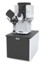
The Helios G4 Plasma Focused Ion Beam CXe Workstation is a fourth generation, fully digital, Extreme High Resolution Field Emission Scanning Electron Microscope (FE SEM) equipped with an inductively coupled plasma (ICP) focused ion beam (PFIB).
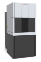
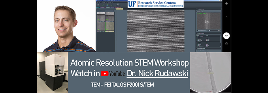
The Talos F200i S/TEM is a flexible and compact 200 kV FEG Scanning Transmission Electron Microscope (S/TEM), which is designed for fast, precise and quantitative characterization of nanomaterials.
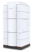
The 300 kV Themis Z is a FEG Scanning Transmission Electron Microscope (S/TEM) with a high-tension voltage range of 60-300 kV.
The Research Service Centers in the Herbert Wertheim College of Engineering are the new home to three state-of-the-art, high-resolution electron microscopes.
The microscopes will provide support and enhance the research, education and public service missions of the University of Florida by providing access to characterization and process instrumentation and facilities. With expert staff on hand to provide assistance and guidance, the students, faculty and industry researchers who use these facilities are ensured the most effective and appropriate use of the center's capabilities.
All of the equipment is scheduled to be available for use and training in Spring of 2020. Announcements on utilization, service and training will be sent to the UF research community upon achieving the installation milestones necessary to start accepting users.

